Thyroid disease is a commonly reported pathology in adult patients. Therefore, the timely diagnosis of pathological processes that have a negative effect on the body is considered a very important point. For this purpose, thyroid gland scintigraphy is used as the most highly informative examination technique.
Material Content:
What is thyroid scintigraphy?
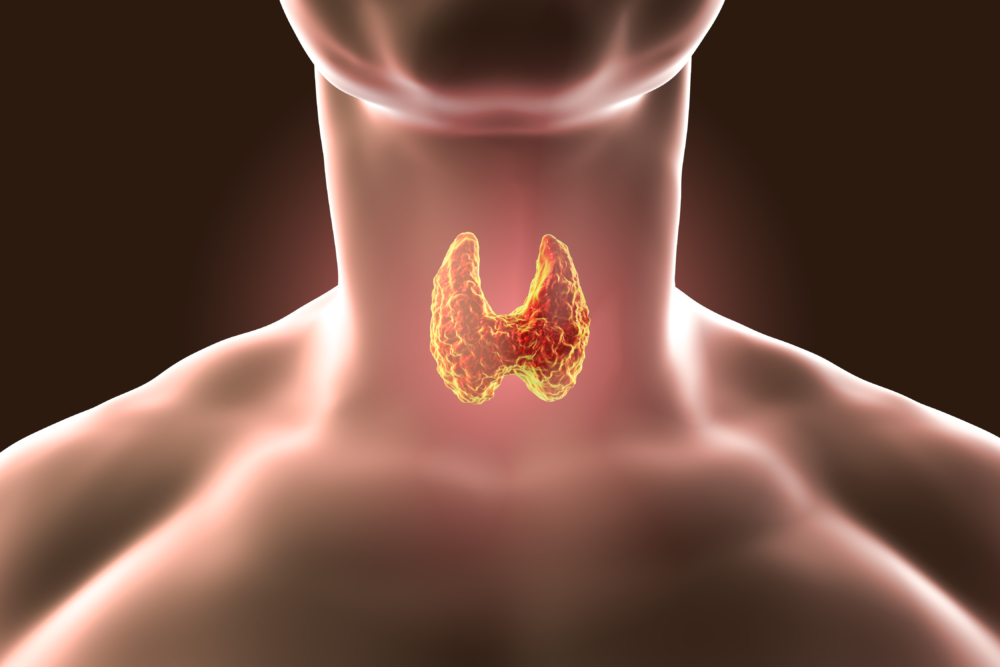
This method of diagnosing diseases is based on the use of radioisotopes, with the help of which a comprehensive examination of the thyroid gland is possible. This organ belongs to the system of endocrine glands, which produces a number of hormones that affect the metabolism, as well as controlling the growth processes of the body.
A feature of the thyroid gland is its ability to absorb, accumulate and remove radioactive isotopes over time. Based on this principle, the diagnosis of gland dysfunction is based, which allows one to determine its shape and size, the presence of nodular formations, pathological changes in the focal or diffuse nature in the structure of the organ.
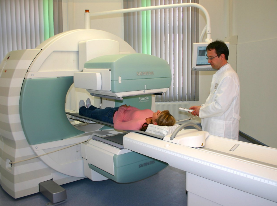
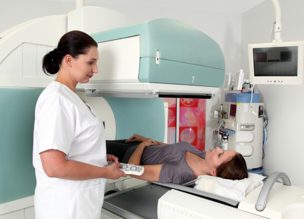
For diagnosis, the radioisotopes of iodine 131 and 123 are used, as well as technetium 99 or an analogue, technetrile 99, which are introduced into the body. Through the bloodstream, these substances spread throughout the body, concentrating more in the thyroid gland.After that, the scanning procedure begins in the gamma camera, the result of which is displayed on the monitor screen.
Indications for the purpose of the study
As a rule, a thyroid gland study by scintigraphy is performed after an ultrasound scan if certain pathologies have been detected and only as directed by the endocrinologist.
The procedure is carried out only for a certain contingent of patients with gland disease, according to the following indications:
- abnormal retrosternal location of the organ;
- pronounced hormonal pathology, not giving the effect of drug treatment;
- abnormal development of the gland with the presence of an anatomical defect;
- inflammatory processes in the organ;
- suspicion of a voluminous process of a malignant nature;
- violation of the functioning of the thyroid gland;
- differentiation of the processes of hormonal activity of organ lobes;
- the presence of thyrotoxicosis.
With caution, the procedure is prescribed for patients prone to allergic reactions.
Examination Preparation and Diet
To obtain a reliable result before the diagnosis, preparation for the examination is necessary, which eliminates the phenomenon of discomfort in the patient.

Activities to be performed before scintigraphy:
- 3 months before the appointed time of the procedure, instrumental examination methods for concomitant diseases using contrast agents (urography, angiography, MRI) are excluded;
- 1-2 months before the scheduled procedure, the administration of drugs containing iodine, as well as thyroid hormones, is stopped;
- external use of an alcoholic solution of iodine is excluded.
Patients receiving medications on an ongoing basis (cardiac, hormonal drugs) should notify the attending physician about this to correct the treatment of concomitant diseases.
Diagnosis with radioisotopes involves a change in diet within a month before the examination. It is necessary to exclude a number of products with a high concentration of iodine from daily use:
- sea fish (cod, pollock, hake);
- fresh herring;
- sea kale;
- shrimp, crabs;
- oysters
- squid;
- greenery;
- persimmon;
- iodine-bromine mineral waters;
- iodized salt.
Women prescribed for diagnosis are recommended to conduct a study in the first half of the menstrual cycle to exclude possible conception.
If thyroid scintigraphy with sodium pertechnetate is performed, preparation for diagnosis is not required. After intravenous administration of the drug, a scan is performed after 30-60 minutes. Slow removal of the drug into the external environment allows you to track the change in the structure of the organ in dynamics.
Important! The appearance of mood swings, a rapid decrease or weight gain, a constant alternation of chills and heat in young women, an unreasonable increase in temperature to subfebrile numbers require an endocrinologist's consultation to exclude thyroid dysfunction.
Diagnostic methods and treatment
Diagnosis of thyroid diseases begins with a general examination of the patient’s organ and its palpation, which makes it possible to assess the shape of the thyroid gland, deviations in its size, the presence of a tumor, nodes, pain on palpation.
After evaluating the objective data, instrumental diagnostic methods are assigned:
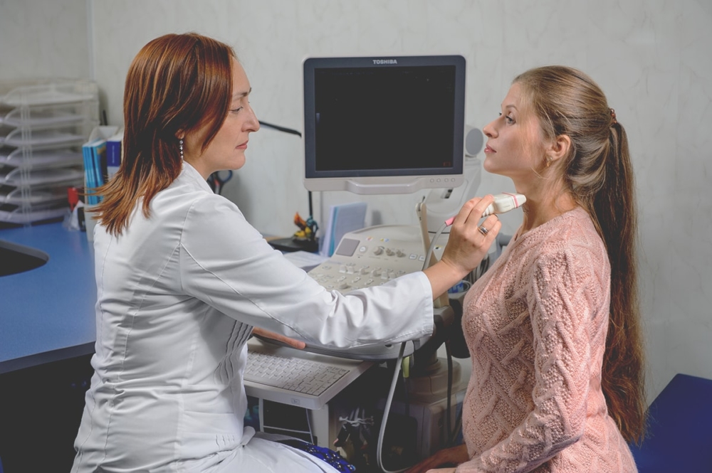
- Ultrasound - using ultrasound, the structure, density of the organ, the presence or absence of nodular formations, and its blood supply are determined. The method allows monitoring the effectiveness of treatment for a malignant process or diagnosing thyrotoxicosis. But not all pathologies are determined by ultrasound;
- biopsy - taking the material for histological examination directly from the densification or node detected by ultrasound;
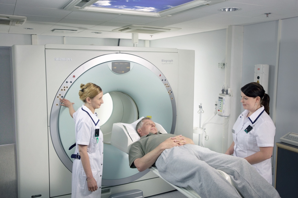
- MRI - is used as an additional diagnostic method for indications, which allows you to study the organ in more detail in its three-dimensional image. This technique is mainly used for retrosternal localization of the thyroid gland;
- laryngoscopy - this study is mainly used before surgery for removal of a gland tumor. The larynx wall is examined to determine the volume of movement of the vocal cords, since a growing tumor can exert pressure on them. This technique allows you to reduce postoperative complications in the vocal cords;
- scintigraphy - performed using radiopharmaceuticals.
After examination and diagnosis, therapeutic measures are prescribed that can be performed conservatively or surgically. The radical method of treatment is used in the case of disease progression with the formation of large nodes and the lack of effect of conservative therapy.
But almost always therapeutic measures begin with the use of radioisotopes. In the treatment of thyroid pathologies, its ability to capture and concentrate all iodine from the body in its structure is used. This property is taken into account during radiotherapy of malignant tumors and endocrine diseases.
Radioactive isotopes
The method of treatment with radioisotopes gives a good effect, stopping the progression of the pathological process in the thyroid gland, without affecting healthy organ cells.
The following thyroid pathologies are an indication for this treatment method:
- diffuse or nodular toxic goiter, developing due to excessive production of thyroid hormones;
- malignant formation.
In the conservative treatment of negative processes in the thyroid gland, a special radioisotope preparation is used.
Iodine
Thyroid radioiodine therapy is a serious procedure that is carried out only in a hospital and has its own characteristics:
- before starting the course of treatment, the patient undergoes hormonal correction in order to increase the absorption capacity of iodine by the cells of the organ;
- a month before therapy, the patient goes on a diet with the abolition of foods rich in iodine;
- in the hospital, the patient is placed in a separate ward, where he takes the capsule with iodine-131 inside. The element, absorbed by the cells of the thyroid gland, destroys the pathological foci in it. This process is monitored during scanning when an assessment is made of the degree of radiation intensity.
After the course of treatment, the patient continues to remain in the hospital for 1-1.5 weeks, being a source of ionizing radiation. After discharge from the hospital, strict adherence to the rules of conduct in everyday life is recommended. Self-medication in this case is excluded.
Thyroid scintigraphy decoding results
Scintigraphy shows the degree of absorption of a radioactive agent with its further distribution in the structure of the gland.
For each nosology, the result of the examination will have its own characteristics:
- norm - uniform accumulation with medium intensity;
- diffuse toxic goiter - uniform accumulation with high intensity;
- hypothyroidism - uniform accumulation with low intensity;
- chronic autoimmune thyroiditis - uneven accumulation;
- toxic adenoma - high absorption in a single area with a clearly defined contour, that is, a "hot" node, practically no absorption is observed in the rest of the tissue;
- nodular goiter - there is no absorption in the area containing the "cold" node;
- thyroid oncology - the presence of nodes with fuzzy borders that unevenly accumulate a radioisotope;
- thyroid cancer metastases - the accumulation of a radioisotope in bones, lymph nodes, lungs, where it should not be.
Based on the results of scintigraphy, a final diagnosis of thyroid disease is made and an appropriate course of treatment is prescribed.
Possible harm and contraindications
This diagnosis is not performed for pregnant women at any time of bearing a child. If it is necessary to carry out scintigraphy of a nursing mother, an examination is done with the radioisotope of technetium, since it is quickly removed from the body along with feces and urine. It will be possible to apply to the baby’s chest one day after the diagnosis.
The method is considered completely safe, since the radioisotopes used for diagnosis are in small quantities and are quickly excreted from the body without causing any harm. Therefore, such a study can be used for children without fear for their health.


