The erythrocyte sedimentation rate (ESR) has been used in laboratory practice since the late 19th and early 20th centuries. Edmund Bernacki, a Polish physician, pathologist and medical historian, proposed using erythrocyte sedimentation as a test. More than 120 years ago, E. Bernatsky published discussions about the possible mechanisms of the phenomenon and observations about the differences in the reaction with different types of pathology. The analysis was named by the author as a erythrocyte sedimentation reaction (RLE). Often when receiving ESR analysis results are higher than normal - what does it mean?
Material Content:
- 1 Normal and pathological indicators of ESR in the blood
- 2 Table norm ESR in women by age
- 3 ESR during pregnancy
- 4 If a woman's ESR level is above normal, what does that mean?
- 5 ESR determination methods
- 6 Diseases in which there is an increased ESR in the blood
- 7 When can an increase in ESR be considered safe?
- 8 How to reduce ESR in the blood?
Normal and pathological indicators of ESR in the blood
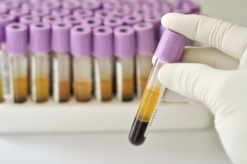
Even in the era of Galen, Hippocrates, doctors actively used bloodletting and noticed that the blood stood "stratified" after standing. The lower layer is more dense and colored, and the upper one is transparent and light. It was noted that in patients the light layer is more pronounced than the dark one. But until the 20th century, the diagnostic value of ESR was not noted.
In 1918, at a congress in Stockholm, the Swedish hematologist R. Fareus announced a change in ESR during gestation, considering this analysis a pregnancy test. Later, ESR was considered an objective test for pathological processes in the body.
The essence of the ESR phenomenon is that red blood cells under the influence of gravity form a precipitate. The rate of their subsidence depends on aggregation (sticking together). In various diseases, red blood cells can form large conglomerates and then ESR increases.
The formation of large conglomerates is due to an increase in:
- fibrinogen and globulin levels;
- plasma viscosity;
- blood cell size.
ESR is affected by:
- analysis method;
- age and gender characteristics.
To obtain discrete results, it is necessary to consider factors affecting the accuracy of the indicator. The table shows the causes of changes in ESR in people, regardless of gender and age:
| Factors of influence on ESR | Fast | Slow |
|---|---|---|
| Red blood cell activity | Anemia | Polycythemia |
| Medication | Oral contraceptives | Non-steroidal analgesics |
| Lipid metabolism | Increase cholesterol | Increased bile acid levels in the blood |
| Violation of the acid-base balance of the blood | Acidosis (“acidification”) | Alkalosis ("alkalization") |
| Ambient temperature during capillary settling | > + 27 ° C | + 22 ° C |
| Other factors | Pregnancy | Anomalies in the size and shape of blood cells |
The influence of these factors distorts the result of the analysis and should be taken into account when conducting research.
ESR can not claim to be the "title" of the analysis, which gives an exhaustive result. When appointing it and deciphering the indicators, the doctor should be aware of the diagnostic limitations of the analysis.
Among women
In women under the age of 60 years, the reference value (norm) of ESR is 2-12 mm / hour. The indicator varies depending on the condition and amount of the main components of the blood, as well as on the activity of androgynous hormones. For both sexes, there are indicators of normal ESR in the blood in accordance with age. So, for women older than 60 years, the norm is considered <20 (30) mm / hour.
A change in the level of hormones is also noted during the gestation period, therefore there is a special table of reference values depending on the gestational age. Adaptation mechanisms in preparation for childbirth include changes in the blood. The rate of ESR in the second half of pregnancy is 40-50 mm / hour.
Since reference values are an average indicator, and the upper limit of the norm is valid only for 95% of patients, individual calculations of the norm can be done using the Tarelli, Westergren or simpler Miller formulas.
In children
The rate of ESR in the blood in children reflects the features of their development and improvement of various functions of the body systems.
So, for example, ESR in the blood of newborns does not exceed 2 mm / hour, which is due to the characteristics of the blood composition:
- high red blood cell count (hematocrit);
- low amount of proteins and, in particular, globulins;
- high cholesterol (hypocholesterolemia);
- low acidosis.
With age, blood counts in children change, and ESR indicators also change.
So, for example, the norm of ESR in children is:
- newborns: 1-7 day - 1-2 mm / hour; 8-14 day - 4-17 mm / hour; 2-6 months - 17-20 mm / hour;
- preschoolers - 1-8 mm / hour;
- adolescents: girls - 15-18 mm / hour; boys - 10-12 mm / hour.
In children, all indicators of the functioning of systems are more labile (mobile) than in adults. Therefore, they are greatly influenced by external factors, such as environmental conditions. It was found that in children and adolescents living in adverse conditions, blood characteristics differ from the average normal values. So, children living in high latitudes (European North) have increased gender (gender) differences in the content of red blood cells.
Compared with adolescents from middle latitudes, they have significant differences in ESR:
- in girls - 6-8 mm / hour (versus 5-6 mm / hour);
- in boys - 6-7 mm / hour (versus 4-5 mm / hour).
In adolescents living in the North, the ESR readings are normally significantly higher than the ESR norm in blood in children of middle latitudes. At the same time, the adaptation of girls to high latitude conditions is more pronounced than that of boys.
In men
The norm of ESR in the blood in men is also subject to age-related metamorphoses:
| Age of men (years) | ESR rate (mm / h) |
|---|---|
| less than 20 | 1,0-10,0 |
| 20-60 | 14,0 |
| >60 | 18,0-35,0 |
Slight - by 1-2 units, an increase in the indicator compared to the norm may indicate attenuation of the pathological process or a violation of the conditions for preparing for the analysis.
With a deviation of the result by 15-30 units, a slight inflammatory process characteristic of colds can be suspected.
An increase or decrease in the indicator by> 30 units indicates a serious process.
An indicator that differs from the norm by 60 or more units, signals serious violations of the state.
Since ESR itself is uninformative and non-specific (it cannot accurately indicate the nature and localization of the pathological process), it is prescribed in combination with other studies.
Table norm ESR in women by age
The chemical and physical composition of blood is influenced by many external and internal factors. Since the female body is more susceptible to changes, including hormonal changes, fluctuations in ESR values depending on age are more pronounced in women than in men.

Age-related changes occurring in the body of a woman can be divided into 5 blocks:
- The formation and development of the body.
- The beginning of puberty.
- Puberty is the childbearing period.
- The beginning of menopause.
- Climax
Each of the blocks is characterized by its own ESR norm, and the period of puberty has a more detailed division. The following table presents the norm of ESR in women by age:
| Age (years) | ESR rate (mm / h) | |
|---|---|---|
| Bottom line | Upper bound | |
| 13 | 1-4 | 12 |
| 13-18 | 3 | 18 |
| 19-30 | 2 | 15 |
| 31-40 | 2 | 20 |
| 41-50 | 0 | 26 |
| 51-60 | 0 | 26 |
| >60 | 2 | 55 |
In addition to the factors listed above that affect the ESR indices, women increase the result of the analysis when the hormonal background changes, which cause:
- menstrual cycle;
- pregnancy;
- postpartum condition and breastfeeding;
- taking oral contraceptives;
- hormone replacement therapy.
The ESR level in women is significantly affected by nutrition. Passion for diets of adolescents and young women leads to a deviation of ESR from the age norm. Significant differences from reference values are noted with an increase or decrease in body mass index.
ESR during pregnancy
Different ESR indicators are observed in women at different gestational periods:
- I trimester - ~ 13-21 mm / hour;
- II trimester - 25 mm / hour;
- III trimester - 30-45 mm / hour.
After childbirth, increased ESR persists for some time (3-4 weeks or more). An increased ROE during pregnancy indicates that the fetus is developing.
If a woman's ESR level is above normal, what does that mean?
Pregnant women have ESR levels above normal, what does this mean? Pregnancy is accompanied by genetically programmed, adaptive processes.
The degree of their severity depends on:
- gestational age;
- number of fruits;
- individual reserve capabilities of a woman’s body.
Even with a physiologically ongoing pregnancy, researchers have noted signs of a systemic inflammation syndrome.
With an increase in gestational age, the ESR indicator also increases, which is due to:
- a decrease in the number of red blood cells due to an increase in the volume of blood plasma;
- an increase in the size of red blood cells;
- increased endogenous intoxication;
- activation of anti-inflammatory cytokines;
- a decrease in the amount of total blood protein;
- an increase in fibrinogen in the blood and an increase in its viscosity.
These adaptive mechanisms lead to accelerated erythrocyte sedimentation.
On the surface of red blood cells, proteins are localized, leading to the aggregation of red blood cells. In addition, the charge on the surface of the membrane of red blood cells also changes. If at the beginning of pregnancy potassium ions predominate, then by the II trimester their level decreases, and in the III trimester sodium ions prevail. By the end of pregnancy, the total accumulation of sodium ions reaches limit values. A change in the charge of the membranes leads to "sticking" of blood cells.
In addition, in pregnant women, lipid metabolism changes.In the blood, the amount of cholesterol, triglycerides and fatty acids, which are the material for the synthesis of steroids necessary for the formation of fetal tissues, increases. All these changes lead to an acceleration of ESR and are the physiological norm during pregnancy. Therefore, ESR during the gestational period loses its diagnostic value of the indicator of the inflammatory process.
But if the ESR significantly exceeds the upper limit of the norm, then this may indicate:
- inflammatory processes in the body;
- infections of the urogenital system;
- pyelonephritis caused by the mechanical effects of a growing fetus;
- late gestosis.
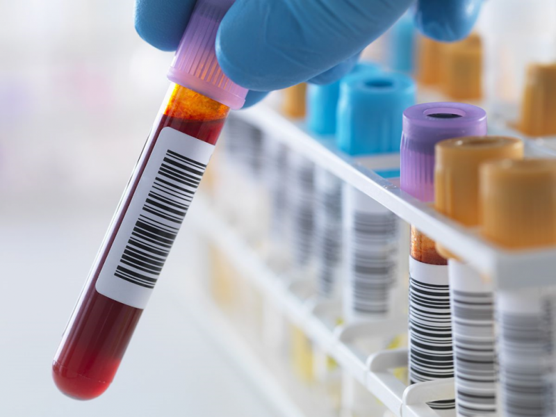
The study of blood parameters, including ESR, is carried out at least 4 times for the entire gestational period. The results obtained help to determine the presence of obstetric complications in the early period and to develop an adequate therapeutic correction of the condition.
ESR determination methods
ESR determination is carried out by several methods. The method proposed in 1924 by T.P. is still popular in our country. Panchenkov. And abroad they use the Westergren method, which in 1977 was recognized by the International Committee for Standardization in Hematology (ICSH) as a standard. The Westergren method was finalized by the Australian hematologist Vintrobe. In Europe and Israel, preference is given to the Vintroba method, and in America, the Vintrobe method is used. How do these methods differ?
ESR by Panchenkov
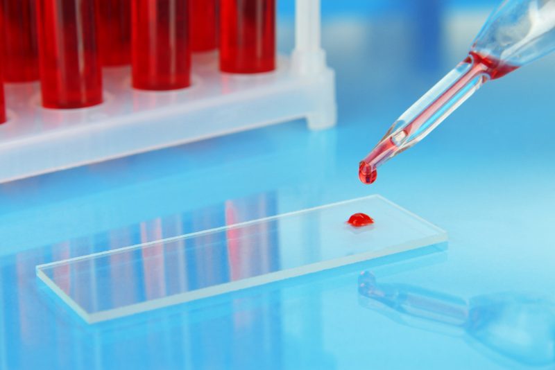
This method has been used for over 90 years to determine ESR. For research, capillary blood is used. It is diluted with sodium citrate and placed in a glass tube with an internal cavity diameter of 1 mm.
The simplicity and cheapness of the method does not compensate for its inherent disadvantages:
- the impossibility of standardizing the method due to the influence of external factors (purity of the capillary, dilution errors, quality of sodium citrate);
- technical difficulties in obtaining capillary blood (hemolysis of blood during compression of the finger);
- the inability to achieve an ideal inner surface and clean capillary during repeated use.
The column used in the analysis is 100 mm long and is graduated in 1 mm increments between risks. Due to the small volume of blood in the capillary, it can not be stored, which is a big drawback when screening. In addition, too many factors affect the accuracy of the result, which does not allow standardizing the method.
ESR for Westergren
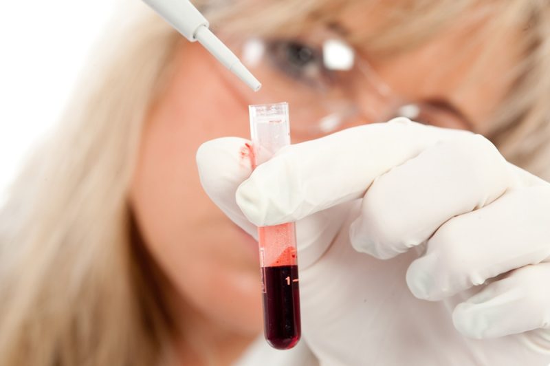
When determining the ROE according to Westergren, whole venous blood is used. The capillary length also differs - it is 200 mm. In the zone of high ESR values in terms of Westergren and Panchenkov, there are differences. So, for example, 70 mm / hour according to Panchenkov corresponds to approximately 100 mm / hour according to Westergren.
Despite the great accuracy of the Westergren method, it also has several disadvantages:
- the inability to use blood for other tests, since blood is prepared differently for general analysis and ESR studies;
- the time is 1 hour;
- high (18.3%) variability of the results;
- the inability to automate the process.
Given these shortcomings, Vintrobe improved the Westergren method.
ESR for Vintrob
According to the Vintrobe method, blood is taken from a vein, but its amount is less than in the Westergren method, since the column is not 200 mm, but 100 mm. But this method greatly underestimates the result, both in the field of low indicators and in the field of high. So, for example, the table shows the corresponding indicators of an ESR blood test, the norm on different scales:
| ESR norm (upper limit) | Westergren (mm / h) | By Vintrobe (mm / h) |
|---|---|---|
| for adult women | 8 | 20 |
| for adult men | 6 | 10 |
| lower bound of unconditioned pathology | 40 | 100 |
Therefore, when indicating ESR indicators, it is necessary to mention which method the study was conducted.
If the Panchenkov and Westergren methods are comparable by results within the normal range, then the Vintrobe method gives indicators that are not comparable with the two previous methods.
In the 90s of the last century, automatic analyzers were developed that translate the result of multiple measurements of the optical density of a blood sample in accordance with the Westergrin scale. This method does not have the disadvantages listed above and the influence of the human factor is excluded.
Diseases in which there is an increased ESR in the blood
Currently, the diagnostic value of the method for determining ESR in various pathological processes is being reviewed.

But for now, the increased ESR value is an indicator of diseases such as:
1. infections caused by various pathological agents:
- bacteria (tuberculosis, infections of the genitourinary system, diseases of the upper and lower respiratory tract);
- viruses (viral hepatitis);
- fungal infections affecting internal organs;
2. malignant diseases:
- malignant blood pathologies;
- malignant neoplasms of various organs;
3. rheumatological diseases (arteritis, rheumatism, rheumatoid arthritis, polymyalgia rheumatism);
4. injuries with suppuration and intoxication;
5. immune diseases and conditions;
6. systemic diseases of the connective tissue (systemic lupus erythematosus, systemic sclerosis, dermatomyositis);
7. pathology of the kidneys (glomerulonephritis, pyelonephritis, renal failure, ICD);
8. endocrine diseases (diabetes mellitus, hyper- or hypofunction of the thyroid gland);
9. other conditions:
- inflammation: gastrointestinal tract, organs of the oral cavity, ENT organs, pelvis, veins of the lower extremities;
- conditions after surgery;
- anemia;
- sarcoidosis;
- allergic reaction;
- epilepsy.
But increased ESR is not always a symptom of a pathological process.
When can an increase in ESR be considered safe?
In addition to the physical and physiological factors listed above that affect the ESR rate, indicators can distort:
- human factor (error or incompetence of the laboratory assistant);
- use of low-quality reagents;
- non-compliance with the rules of preparation for analysis:
- food intake before blood sampling;
- intense physical activity;
- taking hormonal or other drugs;
- prolonged violation of the food and drinking regime (fasting, strict diets, dehydration);
- a change in the gas and lipid composition of the blood.

In children, elevated ESR is observed with:
- lack of vitamins;
- teething;
- a change in nutrition, for example, when switching from breastfeeding to complementary foods;
- malnutrition.
These factors are easily corrected and they do not have a pathogenic effect on the body.
How to reduce ESR in the blood?
In order to reduce elevated ESR, it is necessary to establish the cause, detect and cure the pathology. In the absence of symptoms of the disease, it does not make sense to reduce ESR, studies are prescribed that are repeated after a certain period of time. To regularly monitor the level of ESR in the blood, it is necessary to undergo annual studies, and with an increase in the indicator, additional tests and an in-depth study are prescribed.


