Sand is the smallest formation of concentrated mineral salts and proteins (no larger than 0.9 mm), which only begin to appear in the kidneys and have not yet formed a larger structure. A grain of sand and a very fine stone are often collectively called microlith.
The formation of such tiny grains of sand in the kidneys is a manifestation of the first phase of urolithiasis.
Kidney stones and sand differ only in size and structure. Where does kidney sand come from, symptoms in women and how does it affect the body? Read the answers to these questions below.
Material Content:
Causes
Today, urologists have come to the conclusion that a violation of metabolic (metabolic) processes in the body should be considered as the main reason for the appearance of sand in the kidneys. With the development of this abnormal process, especially if it proceeds for a long time, the content of various salts in the blood and urine increases. If their concentration exceeds the norm, small dense particles begin to form - sand, microliths (small pebbles) and then - large stones.

Certain conditions and diseases can lead to metabolic disorders:
- Genetic hereditary predisposition. The features of metabolic processes in close relatives are very similar, and if someone from the family has been diagnosed with urolithiasis, then the patient is more likely to have sand.
- Deficiency and excess of calcium. The active use of bioactive supplements, vitamins with calcium, increases the likelihood of urolithiasis.
- Excess Vitamin D. Since the vitamin is involved in the metabolism of calcium, with its excess in the blood, the concentration of the substance increases.
- The chemical and mineral composition of the fluid entering the body. If the water that a person drinks is “hard”, then the kidneys have to work with a high load, filtering the solution with an excess of salts of phosphorus, magnesium, calcium.
- Uncontrolled consumption of mineral waters.
- An insufficient amount of fluid drunk per day, which contributes to the concentration of salts.
- Chronic pathologies of the urinary organs and digestive system (structural defects, pyelonephritis, glomerulonephritis, urethritis, peptic ulcer, prostate adenoma). These conditions are characterized by congestion in the kidneys, thickening of the urine, which contributes to the retention of urinary salts in the kidneys.
- Anatomical and physiological abnormalities that interfere with the normal outflow of urine, including reflux - backflow of urine into the urinary ducts from the bladder, narrowing of the urinary duct or its excess.
- Pathology of the thyroid gland. As a result of fluctuations in hormone levels, disorders occur in the filtration and urination systems, and salts begin to settle in the renal pelvis.
- Parathyroid gland dysfunctions (hyperparathyroidism) and excessive secretion of parathyroid hormone, leading to an increase in urine calcium salts.
- Abnormal processes in bone tissue (fractures, osteomyelitis, osteoporosis), in which a high concentration of calcium is observed in the urine.
- Oncological processes in the body.
- Long stay in a hot, dry climate zone. Moisture deficiency leads to saturation of blood and urine with trace elements.
- Predisposing working conditions (in hot shops).
- Long-term forced state of low mobility in severe illnesses, fractures.
- Unbalanced food.
Sand in the kidneys is found by both meat lovers and vegetarians. With significant amounts of animal protein (meat) consumed, urine becomes excessively acidic, and vegetarians, on the contrary, are diagnosed with alkalization. In this case, different types of microliths are formed. In addition, the excess of foods contributing to the violation of the acid-base balance (smoked meats, spicy, sour dishes) and excessive salt intake is of great importance.
Symptoms and signs
Depending on the stage of the disease, anatomical features, and concomitant pathologies, the symptoms of sand in the kidneys in women manifest themselves in different ways.
The initial stage of the process, which is characterized by the appearance of small mineral particles in the kidneys, usually does not give pronounced symptoms, in contrast to the signs that appear when sand comes from the kidneys.
Signs of the presence of microliths in the renal pelvis less than 1 mm are invisible until the particles begin to move in the ureter.
How to understand that the abnormal process has already begun?
It is necessary to monitor such manifestations as:
- Discoloration of urine. It becomes cloudy, more saturated dark yellow.
- The appearance of an unpleasant odor in fresh urine.
- A decrease in urine output per day.
- Change in the composition of urine. This symptom is detected only during diagnostic tests of urine (general and biochemical analysis).
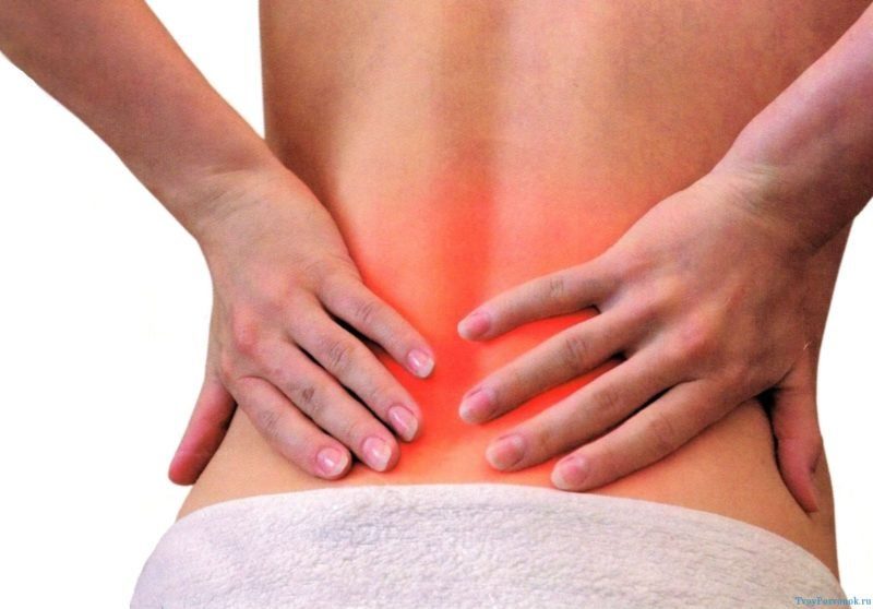
- Sometimes there are slight pulling pains or tingling in the lower back, which go away on their own.
- Fast-passing burning sensation and a very short pain at the end of urination, which most often does not attract attention due to short duration.
- Headaches and frequent increase in blood pressure without diagnosed hypertension and obvious causes such as stress or overwork.
- Symmetrical edema under the eyes in the morning, puffiness.
- Temperature rise. Signals an inflammatory process that began with irritation and damage to the fine tissue of the kidneys with grains of sand.
- Weakness, rapid fatigue.
- Often occurring acute cystitis, vaginal candidiasis, which is associated with activity in the genitourinary tract of pathogenic bacterial flora, fungi against a background of reduced immune defense.
With attention to the symptoms of the first phase of urolithiasis, you can stop the process of stone formation by passing diagnostic tests and starting treatment on time.
Symptoms of the exit of sand from the kidneys in women
Signs of the migration of grains of sand through the ureter are largely reminiscent of symptoms in women with cystitis, appendicitis, inflammation of the appendages and even an ectopic pregnancy. Therefore, one should well understand the features of the clinical picture with sand in the kidneys.

When sand comes from the kidneys, a series of persistent symptoms are noted:
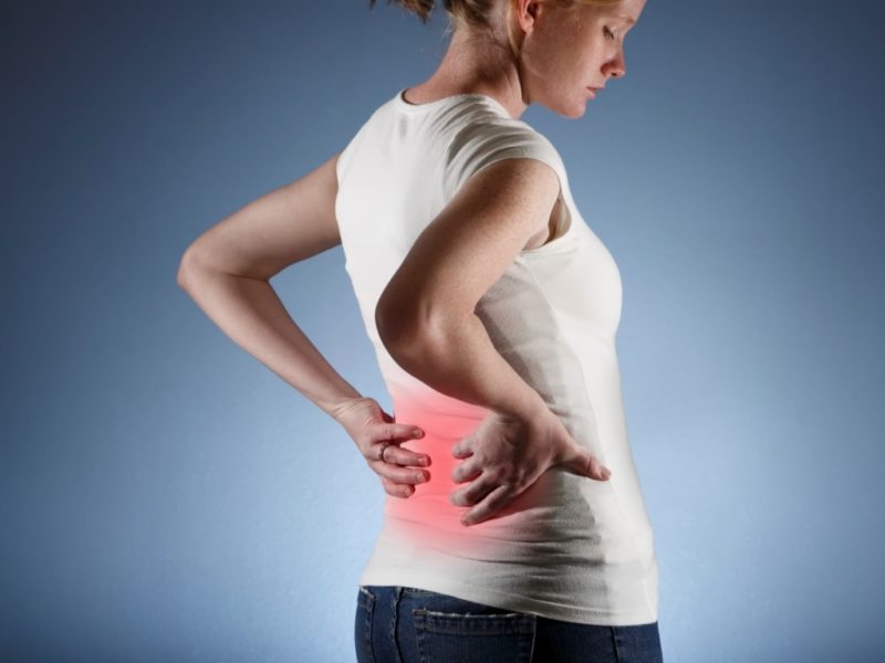
- Drawing or stitching pain in the projection of the urinary ducts - lines stretching down from the navel to the inguinal zone. After the crystals are removed from the urinary tract with urine, an improvement occurs.
- Pain in the lumbar under the ribs. It occurs due to irritation of the walls of the renal pelvis and inflammatory phenomena.
What are the features of pain when moving sand along the urinary tract?
More often pain is associated with physical activity, running, jumping, shaking, which provoke the movement of sand. Painful sensations can occur suddenly, or with a gradual increase, usually from a diseased kidney. But it happens that the whole stomach or lower back hurts. Sometimes the pain radiates (gives) to the groin or intestines. The attack lasts from several hours to 2 days, until the sand comes out when urinating.
Important! It is necessary to distinguish the symptoms of sand output and manifestations with renal colic.
Unlike renal colic, which occurs when the ureter is blocked with a stone of 1 mm or less, pain during the movement of small mineral particles is less acute and can subside, both independently and with medical treatment. This is because grains of sand do not impede the flow of urine and do not block the ureter, as happens with colic.
Pain with renal colic becomes not just severe, but unbearable, which can lead to pain shock.
Urine, in addition to turbidity, acquires a reddish tint due to the presence of blood, since when moving small particles injure the mucous membrane of the urinary tract. Visible particles, mucus, and traces of pus appear in it if purulent inflammation occurs in places damaged by crystals.
- Increased urge to urinate.
- Decrease in daily urine volume.
- Burning, "cramping" and soreness of varying degrees of intensity during emptying of the bladder. Increased pain is noted at the end of urination.
After removing sand with urine, the symptoms of the disease state disappear, but this does not mean that the patient has recovered. The appearance of sand means that the process of stone formation has already begun. Without proper diagnosis and proper treatment, relapse will occur, and urolithiasis will begin to progress, and dense stones will form from the grains of sand.
When to call an ambulance
When sand leaves the urinary tract, the clinical picture can change dramatically if a large calculus begins to move and renal colic occurs. This condition requires immediate treatment.

Therefore, the ambulance crew is called up as soon as the following symptoms appear:
- sudden and very sharp cramping pain, comparable to pain during childbirth with return to the rectum, groin, perineum, stomach;
- very small amount of urine excreted (sometimes dropwise) or inability to urinate at all;
- nausea, vomiting;
- blood clots in the urine visible with the eye;
- imaginary need for bowel movements;
- acute overexcitation, cold sweat;
- rise in blood pressure and body temperature, chills;
- violation of the rhythm and frequency of the heartbeat;
- severe weakness, acute thirst, dry mouth and lips.
It should be understood that often only a few of the symptoms are observed in the patient.
In addition, even without signs of renal colic, emergency care for the release of sand is necessary for children and pregnant women, as the situation can quickly change for the worse.
Diagnostics
To make an accurate diagnosis, the doctor first of all draws attention to the indicated causal factors present in the life of a particular patient, concomitant diseases. The identification of even one of them in combination with characteristic signs gives enough reason to make a preliminary diagnosis.
Lab tests
When conducting general and biochemical analysis, an admixture of protein structures and insoluble salt compounds in the urine that contribute to the formation of sand is determined.

At the same time, the following indicators are analyzed:
- A precipitate of salts (urates, oxalates, phosphates) found in urine indicates the current process of stone formation.
- Wednesday urine. According to this indicator, a predisposition to the formation of a certain type of stones is indirectly determined. Normally, the pH of urine is in the range of 4 - 7. With a slightly acidic environment, oxalates may appear in the kidneys, acidic promotes the formation of urates, alkaline - phosphates.
- The presence of protein above 0.033 g / l indicates an inflammatory process in the kidney tissue.
- The presence of более more than 2 erythrocytes in the field of view ’means that moving sand particles damage the mucous membrane of the ureter, kidney, or urethra and cause bleeding.
- Hyaline cylinders. Their appearance (over 20 in 1 ml) indirectly indicates the formation of calculi, the development of pyelonephritis, hypertension, glomerulonephritis.
- Color. Usually light straw, yellow. A reddish hue indicates the presence of blood.
- Transparency. Cloudy urine happens if mucus, pus, sand is present in it.
- The presence of bacteria. Means infection of the genitourinary tract.
- The presence of white blood cells confirms suspicions of ongoing inflammation. For women and children, the norm is no more than "0 - 6 in sight", for men - "0 - 3".
- Increased urine density is also more often determined with inflammation in the urinary system.
Instrumental diagnostics
Unlike microliths and large calculi, sand in the kidneys is difficult to detect using instrumental diagnostic methods, since the size of the crystals is too small for the equipment to visualize them.
But, although they are more informative in identifying formed calculi, some of them are able to confirm the presence of sand in the renal pelvis or ureter.

The main techniques for diagnosing microliths:
- Ultrasonography. Sometimes it allows you to detect large grains of sand, reveal the presence of stones, as well as inflammatory changes characteristic of the pathology.
- Survey urography or radiography. Allows you to establish anatomical changes in the organs of the genitourinary system, including tumors, narrowing and curvature of the ureters, to detect stones.
- Radioisotope scintigraphy of the kidneys. It is a radionuclide scan in which a radioactive substance that is safe for humans is injected into a vein, so that in the pictures you can see sand in the kidneys.
- Excretory urography of the kidneys. It is considered the most reliable of the techniques for detecting sand in the initial stage of formation. A type of X-ray in which a contrast iodine-containing solution is injected into a vein.
Pregnancy Risk Factors
Many pathological conditions in women awaiting a child are associated with impaired functioning of the urinary system, and the presence of microliths in the kidneys only exacerbates the negative manifestations.
Women in the 3rd trimester of gestation are especially vulnerable.
During this period, processes occur that create favorable conditions for the formation of sand and stones:
- the growing uterus presses the kidneys and urinary tract more and more, forcing them to function in a stressful mode;
- both the process of urination, renal filtration, and urine output slows down;
- there is an imbalance in the balance of electrolytes, acids and alkalis in the body.
These causative factors are prerequisites for the development of nephrolithiasis (urolithiasis) in pregnant women, the second most common diagnosis of renal pathology during this period.
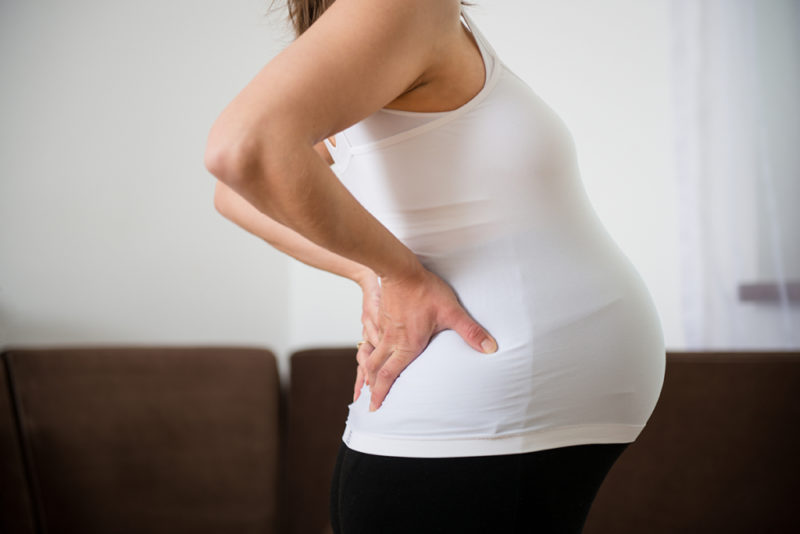
Symptoms in women carrying a child are similar to signs of the presence of mineral particles in the kidneys in all categories of patients. But due to the increased reaction of organs and psyche, they can be more pronounced, painful.
What threat is sand in the kidneys for pregnancy, the health of the expectant mother and the fetus?
Consider the most negative conditions that lead to the formation of mineral renal inclusions:
- Sand and small stones, moving away with urine, are able to partially block the urinary duct squeezed by the uterus, stopping the outflow of urine. This leads to concentration, stagnation of urine and reverse absorption into the blood of poisons, protein breakdown products. Such a process causes a dangerous condition - “urinary tract”, and otherwise poisoning the body (uremia). Such intoxication is dangerous for the development of acute renal failure, fetal death, and termination of pregnancy.
- The release of sand in a pregnant woman can be a harbinger of the movement of a large stone and the further development of renal colic, which is always considered a life-threatening condition.
- Pain in the movement of sand causes the mother to severe stress, vasospasm, which threatens the violation of the uteroplacental circulation and impaired normal development of the fetus.
- The increase in blood pressure, characteristic of the process of migration of grains of sand through the urinary ducts, is also dangerous for the course of pregnancy, since it can lead to placental abruption, impaired blood supply to the baby's organs.
- Stones with a tuberous, spiky surface (often oxalates) damage the tissues of the renal pelvis and ureters, cause constant bleeding, contributing to the introduction of infection and bacterial inflammation of organs.
- Acute pain and inflammation lead to the need for painkillers, antibiotics, the most effective of which, as a rule, are contraindicated during pregnancy, can cause early childbirth or are dangerous for the development of the baby.
Prevention
It is interesting: Ultrasound of the kidneys - preparation for the study
Important! Not all described preventive measures are acceptable for urolithiasis, if there is a large stone in addition to the sand in the organ. The same applies to the state of pregnancy.
If diagnostic tests (ultrasound, excretory urography) prove that there are no stones in the kidneys, then the following measures are recommended to prevent the formation of small crystals:
- Increased daily fluid intake - up to 3 liters (if there are no contraindications): compotes, diluted juices, decoctions of herbs, dried fruits, different varieties of weak tea. The main thing is without preservatives.
- Moderate physical activity running, swimming, skiing, physical exercises to strengthen the pelvic muscles, hips, abdomen, activating blood flow in the urinary organs.
- Diuretic herbal remedies. Contribute to leaching of sand, but with stones can provoke renal colic. Among the main tools are used: Phytolite, Uronefron, Phytolysin, Kanefron, Kidney tea, Urolesan, decoctions of horsetail, onion husks, birch and parsley leaves, lovage root, lingonberry leaf.
- Diet. The appearance in the urine of specific types of salts is often associated with the active consumption of certain products that provoke the development of urolithiasis. The concentration of such salts can be easily reduced by adjusting nutrition.
Read also: horsetail: useful properties and indications
If oxalates are detected, they minimize the amount of products with oxalic acid: citruses, tomatoes, lettuce, sauerkraut, sorrel, strong cocoa, tea and coffee, strawberries, chocolate, spinach, limit milk, salted cheeses. Recommended daily intake of magnesium carbonate, binding salts of oxalic acid.
With urate (uric acid) they reduce the consumption of offal, meat, fried foods, smoked meats, broths, oily fish, mushrooms, vegetable oil, spices, legumes, beer, red wine, radish.
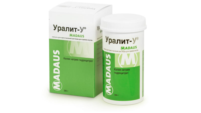
It is advisable to take fresh solutions of citrates (Uralit, Allopurinol, Magurlit, Blemaren), which prevent the formation of uric acid salts and dissolve them.
With phosphate and struvite compounds, alkalization of urine usually occurs, so it needs to be acidified. To do this, minimize the consumption of milk, cottage cheese, cheeses, vegetables, fruits, and increase the number of vegetable oils, meat, eggs and fish.
There is an opinion (not confirmed) that grapefruit juice is generally not advisable to drink with urolithiasis.
Conclusion
If the presence of sand in the kidneys was confirmed during the examination, most patients have enough to recover from a comprehensive conservative treatment, including physiotherapy and diet. There are medicines that promote the resorption of not only sand, but even medium calculi and at the same time normalize the process of urination.


