Osteoscintigraphy is one of the relatively new methods for studying the skeletal system of the human body using the so-called contrasts.
Material Content:
What is osteoscintigraphy?
Scintigraphy of skeleton bones should detect defective areas of bone tissue, where there has been a serious violation of normal metabolism due to destructive-reparative processes. The basis of the method is the determination of the characteristics of the deposition of a contrasting substance in the skeleton, due to which it is possible to accurately indicate the presence of violations, their exact localization, as well as the probable origin. This all helps to greatly facilitate the diagnosis and determine the choice of optimal treatment tactics.
Under normal conditions, radioactive substances do not accumulate in the tissues. In the presence of pathology, they are absorbed by the body and emit gamma radiation, which is read by a special device.
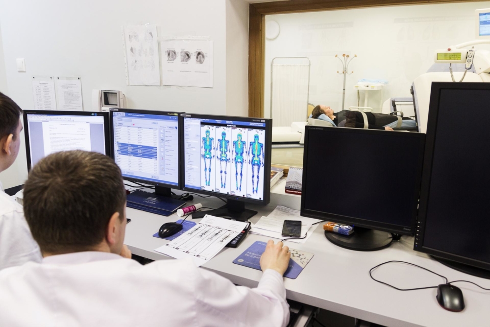
Data is immediately transferred to a computer, on the screen of which the specialist sees a clear image of the organ. Modern cameras allow you to get not only the overall picture, but also to study layered slices.
At its core, the technique is very close to contrast radiography, only in the second case the isotope is injected into the abdominal organ, and not into the bone tissue. However, both research methods identify defects in the accumulation of the isotope, on the basis of which physicians draw conclusions regarding the health and severity of the patient’s disease.
The method is divided into 2 types:
- Dynamic. Take a series of shots without interruption or at regular intervals that show the exact distribution of contrast.
- Static. They take a small number of pictures that simply display a picture of the entire skeleton.
Scanning can be carried out in planar or local mode. In the first case, a full study of the bone tissues of the whole body is carried out, in the second - only specific anatomical sites.
Indications for the purpose of the study
This method is resorted to in such cases:
- Suspicion of oncology or its presence (with prostate cancer, with breast cancer and other organs).
- Extensive metastasis. Diagnosis of bone metastases is most often carried out at the last stages of cancer development. However, it allows you to detect pathological neoplasms 6-7 months earlier than on x-rays. This is a fairly long period of time that can cost a person life.
- Pathological inflammatory processes of bones and joints. Diagnosis of osteomyelitis and rheumatoid arthritis is often carried out in this way.
- The presence of injuries and fractures (especially informative with hidden damage and microcracks).
- The need to differentiate benign and malignant tumors. A biopsy is much more difficult, so most doctors prefer this method.
- Assessment of the overall effectiveness of the prescribed therapy.
- The presence of pain of the musculoskeletal system, the origin of which is not clear.
- Establishing the viability of bone grafts or assessing the degree of rejection.
Which of the research varieties should be preferred, the doctor decides.
Oncology examination preparation and implementation
Special specialized preparation for the examination is not needed. Before the procedure, you can easily have breakfast, interrupting the planned course of treatment is not recommended.
It is advisable to bring with you data from other studies and extracts from treatment sites. All this will help to specify the problem.
Technique of the procedure:
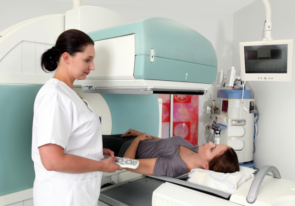
- The introduction of a radioactive substance inside by injection.
- Waiting for 1.5-3 hours (this time is necessary for the isotope to spread throughout the body). It is recommended to wait at the institution and not to go anywhere, as there are cases of an allergic reaction when medical attention is urgently needed.
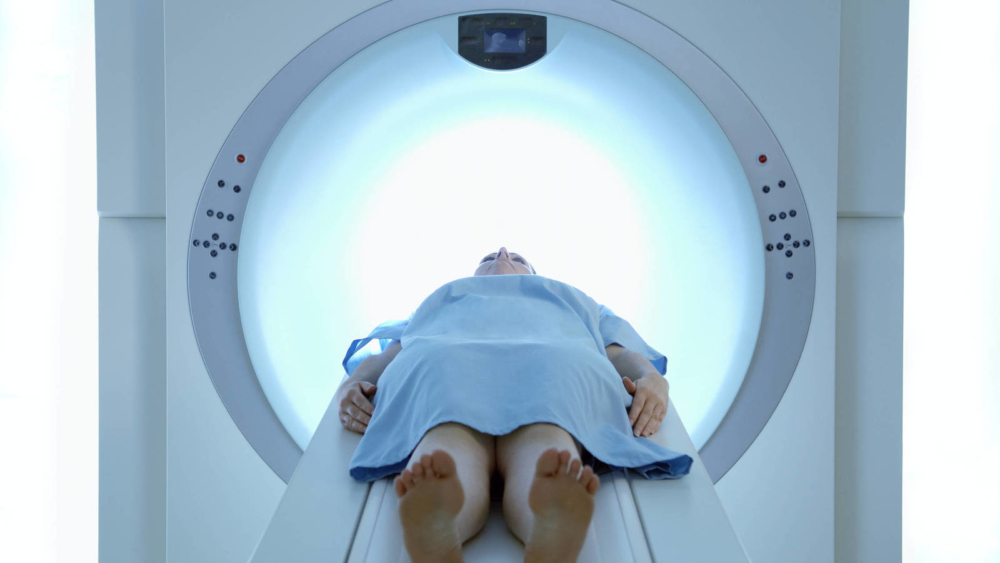
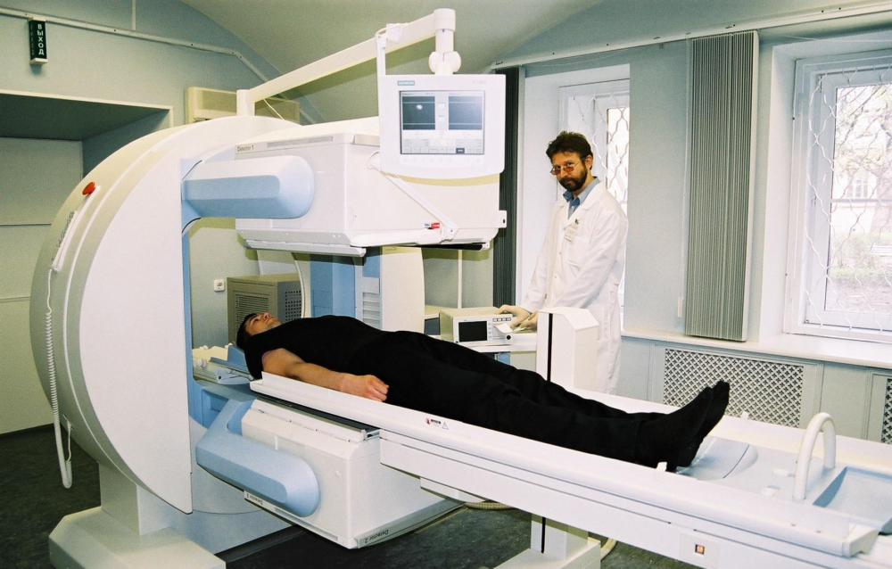
- Performing a scan. The duration of the procedure is from 20 minutes to an hour. It depends on the severity of the disease, the individual characteristics of the body and the number of necessary projections. As a rule, the patient is placed on a special bed on his back, where he should lie still. If this is not possible (for example, a person suffers from severe pain, neurological disorders, or is it a small child who does not want or cannot lie quietly), anesthesia is indicated. No need to undress.
- Waiting for a result.
Within 2 days after the procedure, you need to drink plenty of water in order to quickly remove radiopharmaceuticals from the body. During this period, it is recommended not to particularly contact with other people (especially young children and pregnant women), since there is the possibility of radiation.
In places of pathology, the presence of spots is determined. Bright spots are called "hot" and most often indicate the appearance of metastases. Dull or so-called "cold" points are determined during dystrophic processes of the musculoskeletal system.
Interpretation of the results is carried out by the attending physician. Evaluation of scintigraphy data together with information obtained during CT or MRI allows an accurate diagnosis to be made. However, sometimes for accurate informative data a biopsy specimen is also required, which helps determine the nature of the pathological process.
Methods for scintigraphy of skeleton bones
The sensitivity and high accuracy of the method is based on the ability to detect functional changes.
Radiopharmaceuticals
Only phosphate complexes marked 99mTc are now used to study bone tissue. They have the ability not only to accumulate in metastases, but also in the foci of bone-degenerative lesions of the skeleton. Preparations are prepared right before their introduction in the diagnostic department.
The radiation load on the organs in each case is different, however, it does not exceed the norm.
Isotopic bone examination
Radioisotopes are introduced into the patient's body - special chemicals, the distribution and decay of which are observed in the form of graphic visualization. This helps even in the early stages to recognize bone and joint changes of both a degenerative and inflammatory nature.
Radioactive isotopes are eliminated quite quickly and do not have a harmful effect on health.
Possible complications and contraindications
Of the contraindications, the following should be mentioned:
- the period of bearing a child;
- lactation;
- general serious health condition;
- allergy to the injected substance.
If a nursing woman, according to indications, needs to conduct this study, she needs to temporarily stop breastfeeding. And also after the administration of the drug, an unforeseen reaction of the body may occur, therefore it is impossible to leave the medical institution. Sometimes at the injection site, redness of the skin and burning are observed.
Children's age is not a contraindication. The only caveat - the dose of the drug administered is calculated in accordance with the age and weight of the small patient.
Radiation in comparison with other research methods is not so great. Radiation scintigraphy has important advantages - a very high information content and at the same time minimal harm to the body. The accuracy of the method approaches 90%, although in very rare cases it is possible to obtain a false negative result.


